Two specimens come from Petrified Forest National Park in Arizona, the third from Crosby County in Texas. No single feature can prove that such an ancient fragment truly contains cartilage. Only several clues together expose the fragments for what they are.
A team led by Denycè S. E. Velin of Virginia Tech reports the finding in the journal Palaeodiversity.
The Skeleton That Dissolves Itself
Sharks and their relatives, the cartilaginous fishes, build their internal skeleton from cartilage. Parts of it calcify, meaning they accumulate minerals and become hard. This calcified cartilage is the defining feature of the entire group.
The material has a fatal weakness. It breaks down easily and therefore rarely survives as a fossil. This leaves a gap in the history of shark skeletons that researchers have struggled to close.
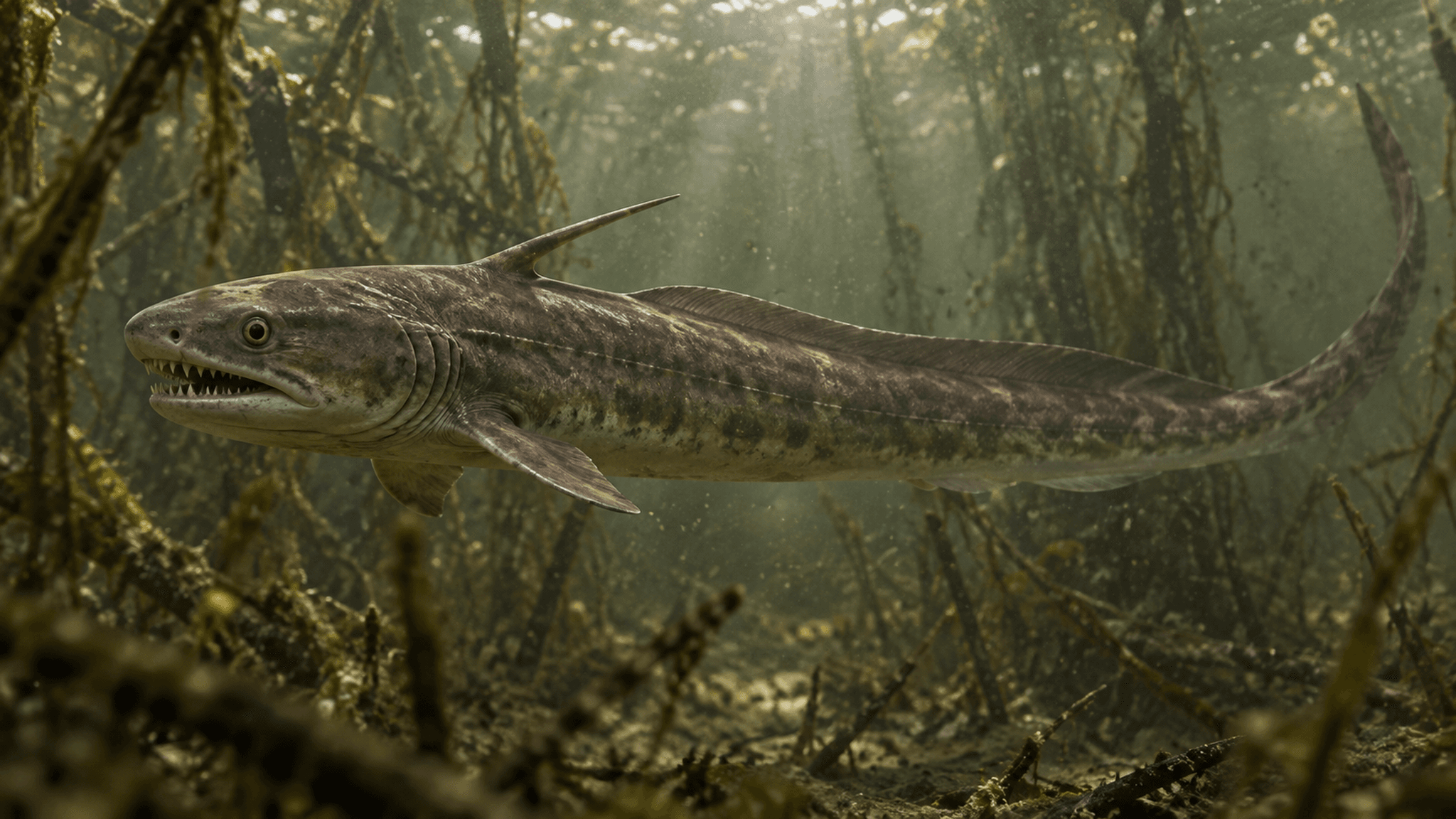
This is how events may have unfolded in the Triassic: a shark dies in freshwater and sinks to the muddy bottom. Currents and decay pull the body apart piece by piece. With luck, a few hard fragments remain and sediment buries them.
The two Arizona specimens lay in the Blue Mesa Member of the Chinle Formation, a rock layer from the early Norian. The Texan specimen comes from the Tecovas Formation. Both layers are around 220 million years old.
A Tiled Mosaic Exposes the Cartilage
The researchers corner the fragments with three tools. Paper-thin slices of rock go under a microscope, while a scanning electron microscope pushes the magnification much further. Computed tomography, or X-rays taken in many layers, probes the interior.
Then the evidence begins to stack up. Polygonal mineral tiles called tesserae sit tightly packed like floor tiles. Fibrous zones connect the mosaic, with tiny cavities between them that specialists call lacunae.
There is no medullary cavity either, the hollow space inside many bones. Each of these traces alone would be only a clue. Together, they form precisely the pattern typical of tessellated calcified cartilage.
No Two Fragments Look Alike
Inside, the finds reveal different faces. In some places, the cartilage calcifies into tiny spheres; elsewhere, it forms column-like structures. Some fragments carry a distinct covering layer above the main body, while others lack this division.
That diversity is exactly what makes the finds valuable. It shows how varied fossil cartilage can look. Anyone who discovers a suspicious fragment in future now has reference images for comparison.
The Trail Leads to Triassic Freshwater Sharks
Which animal carried the cartilage becomes clearer through comparison with living and extinct cartilaginous fishes. The evidence points towards the Xenacanthiformes, a group of sharks. The fragments probably came from such animals.
The locations fit. These sharks are already known from the same sites in both Arizona and Texas. The scene and the suspect therefore match.
Unremarkable Crumbs Become Evidence
The study hits hardest when it turns to material that has been easy to overlook. In fossil beds packed with tiny vertebrate remains, unidentified fragments often lie among teeth and splinters of bone. The tiled mosaic, fibrous zones and internal structure now provide a standard for identifying such crumbs specifically as cartilage.
The three finds therefore close a gap in the fossil record of cartilaginous fishes. They reveal more precisely how their calcified cartilage was built. They also expand the documented evidence for the internal skeletal tissue of sharks from Triassic freshwater environments.
The cartilage of these sharks should have vanished without a trace. Yet three fragments preserved its mosaic for 220 million years, and researchers can now read it.
---